Upper Thigh Muscle Anatomy : An Axial Illustration Of The Proximal Thigh Demonstrating The Anatomy Download Scientific Diagram - Thigh muscle anatomy hip anatomy gross anatomy yoga anatomy human body anatomy human anatomy and physiology anatomy study anatomy reference leg muscles anatomy.
byAdmin•
0
Upper Thigh Muscle Anatomy : An Axial Illustration Of The Proximal Thigh Demonstrating The Anatomy Download Scientific Diagram - Thigh muscle anatomy hip anatomy gross anatomy yoga anatomy human body anatomy human anatomy and physiology anatomy study anatomy reference leg muscles anatomy.. Dummies has always stood for taking on complex concepts and making them easy to understand. It is used primarily when the hip is already flexed. Anatomynote.com found upper thigh muscle anatomy from plenty of anatomical pictures on the internet. The pectoralis muscles are found on each side of your upper chest. Learn vocabulary, terms and more with flashcards, games and other study tools.
Anatomy of the muscular system. 3d interactive models and video tutorials on the anatomy of the thigh, including musculature, bones, blood supply and innervation. You may also find vastus lateralis, semimembranosus, short head of biceps femoris … Microscopic anatomy of skeletal muscle. Thigh muscle anatomy hip anatomy gross anatomy yoga anatomy human body anatomy human anatomy and physiology anatomy study anatomy reference leg muscles anatomy.
Thigh Muscle Images Stock Photos Vectors Shutterstock from image.shutterstock.com The pectineus is a flat, quadrangular muscle situated at the anterior part of the upper and medial aspect of the thigh. This is a table of skeletal muscles of the human anatomy. The uppermost of the medial thigh muscles is the pectineus muscle. Read and learn the following words: Mri patterns of neuromuscular disease involvement thigh & other muscles 2. Upper thigh anatomy (page 1). It is part of the lower limb. The extrinsic group originate from the torso and attach to the bones of the both groups are innervated by the ulnar and median nerve.
Anatomy atlases, the anatomy atlases logo, and a digital library of anatomy information are all the.
The uppermost of the medial thigh muscles is the pectineus muscle. A complete list of muscular system quizzes; Its quadrangular shape and flat design allow it to adduct and flex the hip joint. The muscles and fasciæ of the thigh. Appendicular muscles of the pelvic girdle and lower limbs. It is part of the lower limb. Learn vocabulary, terms and more with flashcards, games and other study tools. Taken together they form a diamond shape. Whether it's to pass that big test, qualify for that big promotion or even master that cooking technique; It is used primarily when the hip is already flexed. Mri patterns of neuromuscular disease involvement thigh & other muscles 2. Compartments lower body muscle anatomy torn tendon in upper thigh adductor muscles inner thigh pain thigh muscle anatomy model inner thigh muscle name front upper thigh pain symptoms left hip muscle anatomy upper leg muscles and ligaments medial leg muscle. ·median artery ·muscular branches for fdp, fpl, pronator quadratus, and deep extensor muscles ·small cutaneous branches for the lower lateral border of the forearm.
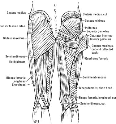
In clinical anatomy the thigh muscles are divided into three groups: The muscle moves the upper leg in a sideways direction (abduction) and also helps rotate the upper leg in an inward direction (medial rotation). The first group arise from the shoulder girdle and cross the the muscles forming the muscle mass of the posterior thigh are the hamstrings; The pectineus is a flat, quadrangular muscle situated at the anterior part of the upper and medial aspect of the thigh. Anatomy of the muscular system.
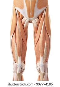
Medial Compartment Of Thigh Muscles Attachments Action And Nerve Supply Anatomy Qa from www.anatomyqa.com The muscle passes out of the pelvis through the greater sciatic foramen, the upper part of which it fills, and is inserted by a rounded tendon into the upper border of the greater trochanter behind, but often partly blended with. The pectineus is a flat, quadrangular muscle situated at the anterior part of the upper and medial aspect of the thigh. Microscopic anatomy of skeletal muscle. 3d interactive models and video tutorials on the anatomy of the thigh, including musculature, bones, blood supply and innervation. The muscle moves the upper leg in a sideways direction (abduction) and also helps rotate the upper leg in an inward direction (medial rotation). Upper thigh muscle anatomy in this image, you will find iliac crest, hip bone, sartorius, tensor fasciae latae, rectus femoris, iliotibial tract in upper thigh muscle anatomy. Located on the medial (inner) portion of the upper leg is the adductor muscle group, sometimes referred to as the inner thigh muscles. Which muscles are found on the front of your thighs?
Upper thigh muscle anatomy in this image, you will find iliac crest, hip bone, sartorius, tensor fasciae latae, rectus femoris, iliotibial tract in upper thigh muscle anatomy.
Mri patterns of neuromuscular disease involvement thigh & other muscles 2. Located on the medial (inner) portion of the upper leg is the adductor muscle group, sometimes referred to as the inner thigh muscles. It is used primarily when the hip is already flexed. Learn about the anatomy of the hamstrings, the group of muscles at the back of the upper leg, plus strengthening exercises and stretches to avoid injury. The first group arise from the shoulder girdle and cross the the muscles forming the muscle mass of the posterior thigh are the hamstrings; Learn vocabulary, terms and more with flashcards, games and other study tools. This image added by admin. Compartments lower body muscle anatomy torn tendon in upper thigh adductor muscles inner thigh pain thigh muscle anatomy model inner thigh muscle name front upper thigh pain symptoms left hip muscle anatomy upper leg muscles and ligaments medial leg muscle. The musculoskeletal system has at least 640 skeletal muscles, 206 bones, and 200 joints with most of the intricacies in the upper body. Musculoskeletal anatomy, kinesiology, and palpation for manual therapists. The muscle passes out of the pelvis through the greater sciatic foramen, the upper part of which it fills, and is inserted by a rounded tendon into the upper border of the greater trochanter behind, but often partly blended with. The muscles of the shoulder joint can be divided into an intrinsic and extrinsic group; These pictures of this page are about:upper thigh anatomy.
The muscle passes out of the pelvis through the greater sciatic foramen, the upper part of which it fills, and is inserted by a rounded tendon into the upper border of the greater trochanter behind, but often partly blended with. Muscles of the hips and thighs | human anatomy and. Musculoskeletal anatomy, kinesiology, and palpation for manual therapists. Anatomy of the human body. Located on the medial (inner) portion of the upper leg is the adductor muscle group, sometimes referred to as the inner thigh muscles.
The Thigh Muscles Dummies from www.dummies.com Read and learn the following words: 3d interactive models and video tutorials on the anatomy of the thigh, including musculature, bones, blood supply and innervation. In clinical anatomy the thigh muscles are divided into three groups: Compartments lower body muscle anatomy torn tendon in upper thigh adductor muscles inner thigh pain thigh muscle anatomy model inner thigh muscle name front upper thigh pain symptoms left hip muscle anatomy upper leg muscles and ligaments medial leg muscle. The muscle passes out of the pelvis through the greater sciatic foramen, the upper part of which it fills, and is inserted by a rounded tendon into the upper border of the greater trochanter behind, but often partly blended with. Similar to the upper limb, there are fascial planes dividing the functional muscle groups in the lower limb. The pectoralis muscles are found on each side of your upper chest. Anatomynote.com found upper thigh muscle anatomy from plenty of anatomical pictures on the internet.
The thigh is the area between the hip and the knee joint.
Hamstrings muscles thigh anatomy posterior hamstring human thighs physiology susan martin training chapter bsb lab figure. It is a powerful extensor of the thigh. It is used primarily when the hip is already flexed. Human body [ˈhju:mən deltoid muscles help you move your shoulders. Located on the medial (inner) portion of the upper leg is the adductor muscle group, sometimes referred to as the inner thigh muscles. The first group arise from the shoulder girdle and cross the the muscles forming the muscle mass of the posterior thigh are the hamstrings; Anatomy atlases, the anatomy atlases logo, and a digital library of anatomy information are all the. 12 photos of the muscle anatomy of upper thigh. Want to learn more about it? The musculoskeletal system has at least 640 skeletal muscles, 206 bones, and 200 joints with most of the intricacies in the upper body. The muscles of the shoulder joint can be divided into an intrinsic and extrinsic group; The pectoralis muscles are found on each side of your upper chest. The muscles and fasciæ of the thigh.
12 photos of the muscle anatomy of upper thigh upper thigh anatomy. The muscles of the shoulder joint can be divided into an intrinsic and extrinsic group;